
")


Fax :+39064440062
andrea.ilari@cnr.it
Director of Research
Istitute of Molecular Biology and Pathology - National Research Council
Department of Biochemical Sciences -A. Rossi Fanelli.
Sapienza University of Rome - Edificio di Chimica Biologica Piano terra, stanza T10 P.le A. Moro 5 - 00185 Roma
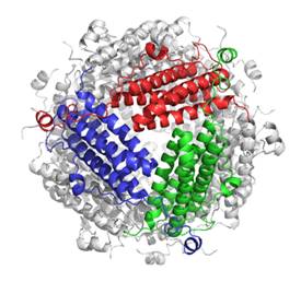
1:Studies on structure-function relationships in cage-like proteins involved in mechanism of defence from oxidative damage Since the early years of my scientific career I have been concerned with defence mechanisms against oxidative damage. In particular, in 2000 I began studying a class of proteins produced by bacteria in starvation conditions that protect DNA from oxidative damage: Dps (DNA binding proteins from starved cells). I solved the X-ray structure of the Dps of Listeria innocua in 2000, the work reporting the structural analysis of the Dps of Listeria innocua (1) has aroused considerable interest in the scientific community and has been cited for this 220 times, as it is a paradigmatic example of how the structure allows us to understand the protein function. In particular, the structure of the protein made it possible to discover that Dps are able to oxidize and incorporate iron into the internal cavity and the oxidation of iron occurs thanks to a ferroxidase center present at the interface of two subunits. These studies were financed by the CNR with the Young Researchers project (2000-2002). Subsequent work on the Dps of other bacteria has made it possible to understand that ferroxidase activity is one of the mechanisms protecting DNA from oxidative damage, since through this activity the hydrogen peroxide produced during oxidative metabolism is consumed (2). Structural studies have made it possible to understand that the protein is made up of 12 identical subunits assembled with 23 symmetry to form a hollow sphere in which iron (up to 500 ions) is stored in microcrystalline form (3). Dps is therefore capable of forming ferric oxyhydroxide nanoparticles and this property is very important from a biotechnological point of view. Both Dps and Ferritins (cage-like proteins homologous to Dps but which form hollow spheres made up of 24 subunits) are capable of synthesizing nanoparticles of other metals. The mechanism by which ferritins synthesize silver nanoparticles was described by my studies on the ferritin of Pyrococcus furiosus, published in JACS (4) and (cited 100 times). References
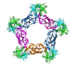
2:Sorcin: a protein involved in calcium mediated signal transduction and multidrug resistance Another topic I have dealt with is calcium-mediated signal transduction. In particular, since the first years of my activity I have been dealing with the structure-activity relationship in Sorcin (Soluble Resistance related Calcium bInding proteiN), a protein that is overexpressed in various tumor lines resistant to chemotherapy and which is involved both in calcium-mediated signal transduction than in “multidrug resistance”. In 2002 I solved the high resolution structure of the SCBD (Sorcin Calcium Binding Domain) (5) which demonstrated that Sorcin belongs to the PEF family (Penta-EF Hands calcium binding proteins) and that the calcium binding with determines a conformational variation that exposes hydrophobic surfaces which make it capable of interacting with its molecular partners. Subsequent studies in which I participated have highlighted that sorcin is able to interact with a series of calcium channels: the ryanodine receptor (RYR2), the sodium calcium exchanger (NCX1), the voltage-dependent calcium channel (LTCC), and the sarcoplasmic and endoplasmic calcium ATPase (SERCA), thus regulating calcium homeostasis. Thanks to the resolution of the structures of calcium-bound and apo human sorcin, we have discovered the molecular mechanism by which the conformational variation induced by calcium promotes the interaction with its molecular partners (6). The importance of Sorcin in preventing stress of the endoplasmic reticulum and regulating the flow of calcium between ER and mitochondria through the MAM (mitochondria-associated mambrane) is of fundamental importance, even in neurodegenerative diseases such as HD, PD, AD. In fact, in cellular models of these diseases sorcin is overexpressed to counteract metabolic imbalances caused by endoplasmic reticulum stress as I demonstrated together with my collaborators (7). Finally, I contributed to demonstrating with my research that sorcin is overexpressed in many types of cancer and is significantly upregulated in cell lines resistant to different chemotherapeutics (MultiDrug Resistant). Thanks to my contribution it was discovered that sorcin is able to bind doxorubicin and other chemotherapeutics and is therefore expressed in large quantities in MDR cancer cells (8). References
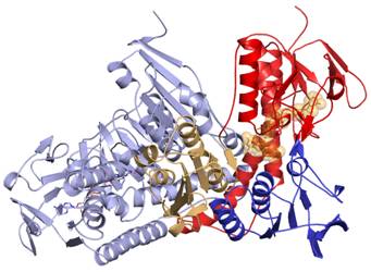
3: Targeting the trypanothione metabolism enzymes good target to find new drugs against Leishmaniasis
Since 2008 I have become interested in Leishmaniasis, a disease transmitted by sandflies which affects populations in tropical and subtropical areas in America, Asia and Africa but is also spreading in southern Europe. It is a disease that if left untreated can lead to death and in fact kills 60,000 people a year. Leishmaniasis is a neglected disease because it affects the poorest part of the world's population. Leishmania enters the host's blood as a promastigote following the bite of the sandfly and is phagocytosed by macrophages where it differentiates into an amastigote and multiplies. The reactive oxygen species produced by macrophages are neutralized by the parasite thanks to trypanothione, a molecule present in large quantities in the protozoan. The trypanothione metabolism enzymes are therefore essential enzymes for the survival of Leishmania and trypanosomatids in general and, being absent in humans, they are good targets for finding new "lead compounds" against leishmaniasis. The first contribution I made in this field of research concerned the solution of the crystal structure of Trypanothione Reductase (TR) and the other enzymes of the trypanothione metabolism (9,10). At the same time, I studied the mechanism of action of antimonials, the most used drugs against this disease, and I discovered that antimony (III), the active ingredient, is able to bind to two catalytic cysteines of TR, thus blocking the reduction of trypanothione and therefore its activation (10). In the following years I then concentrated on "Structure.Based Drug design" (SBDD) and thanks to the collaboration with various groups of pharmaceutical chemists from public and private institutions, I contributed to the design of inhibitors capable of inhibiting TR at a submicromolar concentration and therefore killing the parasite without being toxic to humans (11, 12). References
4:Structure-function relationship in proteins involved in molecular mechamism linked to Huntington Disease.
Lately I have begun to deal with the molecular mechanisms linked to Huntington's disease (HD). Huntington's disease is a rare autosomal dominant genetic disease caused by the mutation of the Huntingtin gene where an expansion of the poly-CAG tract (which corresponds to a polyglutamine tract) occurs. The mutated protein (mHtt) (which contains a stretch of polyglutamines greater than 35 units) causes imbalances in energy metabolism and calcium metabolism in neuronal cells. The toxicity of mHtt is increased by the sumoylation process which makes the protein soluble and makes it capable of migrating into the nucleus and therefore binding to different targets, thus altering cellular metabolism. Sumoylation is catalyzed by the Rhes protein (Ras Homolog Enriched in Striatum). Thanks to the studies conducted by me and my collaborators it was possible to create a model of Rhes (Ras Homolog Enriched in Striatum) and hypothesize the mHtt sumoylation mechanism which could be useful for the design of peptides that inhibit this process (14). Another interesting protein for its involvement in HD and other neurodegenerative diseases is the sigma1 receptor, an endoplasmic receptor involved in repair mechanisms (Unfolding Protein Response), in the regulation of autophagy and in the regulation of calcium flows from the endoplasmic reticulum to the mitochondria. Our studies funded by the Ministry of Health (finalized research 2016, RF 2016-02364123) have contributed to revealing the working mechanisms of S1R. We found that S1R interacts with Sorcin and this may be one of the mechanisms by which the protein controls calcium fluxes from the ER to the mitochondrion via the MAMs (7). Since the activation of S1R has as its ultimate consequence the protection of the neuron from damage caused by protein aggregation, my studies have focused on the search for new S1R agonists through SBDD. In particular, applying the concept of "Drug repurposing" we carried out the VS of the "FDA-approved drugs" on the structure of S1R by selecting some molecules which through cell biology assays we have demonstrated to be receptor agonists (15). Among them the most promising selected S1R agonist appear to be Iloperidone an atypical antipsychotics. Finally, our studies contributed at identifying the compounds with steroid scaffold as preferred ligands for S1R and at discovering how the ligands may reach the binding site i.e through the lipidic membrane. (16). References
|
1:Studies on structure-function relationships in cage-like proteins involved in mechanism of defence from oxidative damage Since the early years of my scientific career I have been concerned with defence mechanisms against oxidative damage. In particular, in 2000 I began studying a class of proteins produced by bacteria in starvation conditions that protect DNA from oxidative damage: Dps (DNA binding proteins from starved cells). I solved the X-ray structure of the Dps of Listeria innocua in 2000, the work reporting the structural analysis of the Dps of Listeria innocua (1) has aroused considerable interest in the scientific community and has been cited for this 220 times, as it is a paradigmatic example of how the structure allows us to understand the protein function. In particular, the structure of the protein made it possible to discover that Dps are able to oxidize and incorporate iron into the internal cavity and the oxidation of iron occurs thanks to a ferroxidase center present at the interface of two subunits. These studies were financed by the CNR with the Young Researchers project (2000-2002). Subsequent work on the Dps of other bacteria has made it possible to understand that ferroxidase activity is one of the mechanisms protecting DNA from oxidative damage, since through this activity the hydrogen peroxide produced during oxidative metabolism is consumed (2). Structural studies have made it possible to understand that the protein is made up of 12 identical subunits assembled with 23 symmetry to form a hollow sphere in which iron (up to 500 ions) is stored in microcrystalline form (3). Dps is therefore capable of forming ferric oxyhydroxide nanoparticles and this property is very important from a biotechnological point of view. Both Dps and Ferritins (cage-like proteins homologous to Dps but which form hollow spheres made up of 24 subunits) are capable of synthesizing nanoparticles of other metals. The mechanism by which ferritins synthesize silver nanoparticles was described by my studies on the ferritin of Pyrococcus furiosus, published in JACS (4) and (cited 100 times). References
2:Sorcin: a protein involved in calcium mediated signal transduction and multidrug resistance Another topic I have dealt with is calcium-mediated signal transduction. In particular, since the first years of my activity I have been dealing with the structure-activity relationship in Sorcin (Soluble Resistance related Calcium bInding proteiN), a protein that is overexpressed in various tumor lines resistant to chemotherapy and which is involved both in calcium-mediated signal transduction than in “multidrug resistance”. In 2002 I solved the high resolution structure of the SCBD (Sorcin Calcium Binding Domain) (5) which demonstrated that Sorcin belongs to the PEF family (Penta-EF Hands calcium binding proteins) and that the calcium binding with determines a conformational variation that exposes hydrophobic surfaces which make it capable of interacting with its molecular partners. Subsequent studies in which I participated have highlighted that sorcin is able to interact with a series of calcium channels: the ryanodine receptor (RYR2), the sodium calcium exchanger (NCX1), the voltage-dependent calcium channel (LTCC), and the sarcoplasmic and endoplasmic calcium ATPase (SERCA), thus regulating calcium homeostasis. Thanks to the resolution of the structures of calcium-bound and apo human sorcin, we have discovered the molecular mechanism by which the conformational variation induced by calcium promotes the interaction with its molecular partners (6). The importance of Sorcin in preventing stress of the endoplasmic reticulum and regulating the flow of calcium between ER and mitochondria through the MAM (mitochondria-associated mambrane) is of fundamental importance, even in neurodegenerative diseases such as HD, PD, AD. In fact, in cellular models of these diseases sorcin is overexpressed to counteract metabolic imbalances caused by endoplasmic reticulum stress as I demonstrated together with my collaborators (7). Finally, I contributed to demonstrating with my research that sorcin is overexpressed in many types of cancer and is significantly upregulated in cell lines resistant to different chemotherapeutics (MultiDrug Resistant). Thanks to my contribution it was discovered that sorcin is able to bind doxorubicin and other chemotherapeutics and is therefore expressed in large quantities in MDR cancer cells (8). References
3: Targeting the trypanothione metabolism enzymes good target to find new drugs against Leishmaniasis
Since 2008 I have become interested in Leishmaniasis, a disease transmitted by sandflies which affects populations in tropical and subtropical areas in America, Asia and Africa but is also spreading in southern Europe. It is a disease that if left untreated can lead to death and in fact kills 60,000 people a year. Leishmaniasis is a neglected disease because it affects the poorest part of the world's population. Leishmania enters the host's blood as a promastigote following the bite of the sandfly and is phagocytosed by macrophages where it differentiates into an amastigote and multiplies. The reactive oxygen species produced by macrophages are neutralized by the parasite thanks to trypanothione, a molecule present in large quantities in the protozoan. The trypanothione metabolism enzymes are therefore essential enzymes for the survival of Leishmania and trypanosomatids in general and, being absent in humans, they are good targets for finding new "lead compounds" against leishmaniasis. The first contribution I made in this field of research concerned the solution of the crystal structure of Trypanothione Reductase (TR) and the other enzymes of the trypanothione metabolism (9,10). At the same time, I studied the mechanism of action of antimonials, the most used drugs against this disease, and I discovered that antimony (III), the active ingredient, is able to bind to two catalytic cysteines of TR, thus blocking the reduction of trypanothione and therefore its activation (10). In the following years I then concentrated on "Structure.Based Drug design" (SBDD) and thanks to the collaboration with various groups of pharmaceutical chemists from public and private institutions, I contributed to the design of inhibitors capable of inhibiting TR at a submicromolar concentration and therefore killing the parasite without being toxic to humans (11, 12). References
4:Structure-function relationship in proteins involved in molecular mechamism linked to Huntington Disease.
Lately I have begun to deal with the molecular mechanisms linked to Huntington's disease (HD). Huntington's disease is a rare autosomal dominant genetic disease caused by the mutation of the Huntingtin gene where an expansion of the poly-CAG tract (which corresponds to a polyglutamine tract) occurs. The mutated protein (mHtt) (which contains a stretch of polyglutamines greater than 35 units) causes imbalances in energy metabolism and calcium metabolism in neuronal cells. The toxicity of mHtt is increased by the sumoylation process which makes the protein soluble and makes it capable of migrating into the nucleus and therefore binding to different targets, thus altering cellular metabolism. Sumoylation is catalyzed by the Rhes protein (Ras Homolog Enriched in Striatum). Thanks to the studies conducted by me and my collaborators it was possible to create a model of Rhes (Ras Homolog Enriched in Striatum) and hypothesize the mHtt sumoylation mechanism which could be useful for the design of peptides that inhibit this process (14). Another interesting protein for its involvement in HD and other neurodegenerative diseases is the sigma1 receptor, an endoplasmic receptor involved in repair mechanisms (Unfolding Protein Response), in the regulation of autophagy and in the regulation of calcium flows from the endoplasmic reticulum to the mitochondria. Our studies funded by the Ministry of Health (finalized research 2016, RF 2016-02364123) have contributed to revealing the working mechanisms of S1R. We found that S1R interacts with Sorcin and this may be one of the mechanisms by which the protein controls calcium fluxes from the ER to the mitochondrion via the MAMs (7). Since the activation of S1R has as its ultimate consequence the protection of the neuron from damage caused by protein aggregation, my studies have focused on the search for new S1R agonists through SBDD. In particular, applying the concept of "Drug repurposing" we carried out the VS of the "FDA-approved drugs" on the structure of S1R by selecting some molecules which through cell biology assays we have demonstrated to be receptor agonists (15). Among them the most promising selected S1R agonist appear to be Iloperidone an atypical antipsychotics. Finally, our studies contributed at identifying the compounds with steroid scaffold as preferred ligands for S1R and at discovering how the ligands may reach the binding site i.e through the lipidic membrane. (16). References
# A. Ilari is corresponding author |
1:Studies on structure-function relationships in cage-like proteins involved in mechanism of defence from oxidative damage Since the early years of my scientific career I have been concerned with defence mechanisms against oxidative damage. In particular, in 2000 I began studying a class of proteins produced by bacteria in starvation conditions that protect DNA from oxidative damage: Dps (DNA binding proteins from starved cells). I solved the X-ray structure of the Dps of Listeria innocua in 2000, the work reporting the structural analysis of the Dps of Listeria innocua (1) has aroused considerable interest in the scientific community and has been cited for this 220 times, as it is a paradigmatic example of how the structure allows us to understand the protein function. In particular, the structure of the protein made it possible to discover that Dps are able to oxidize and incorporate iron into the internal cavity and the oxidation of iron occurs thanks to a ferroxidase center present at the interface of two subunits. These studies were financed by the CNR with the Young Researchers project (2000-2002). Subsequent work on the Dps of other bacteria has made it possible to understand that ferroxidase activity is one of the mechanisms protecting DNA from oxidative damage, since through this activity the hydrogen peroxide produced during oxidative metabolism is consumed (2). Structural studies have made it possible to understand that the protein is made up of 12 identical subunits assembled with 23 symmetry to form a hollow sphere in which iron (up to 500 ions) is stored in microcrystalline form (3). Dps is therefore capable of forming ferric oxyhydroxide nanoparticles and this property is very important from a biotechnological point of view. Both Dps and Ferritins (cage-like proteins homologous to Dps but which form hollow spheres made up of 24 subunits) are capable of synthesizing nanoparticles of other metals. The mechanism by which ferritins synthesize silver nanoparticles was described by my studies on the ferritin of Pyrococcus furiosus, published in JACS (4) and (cited 100 times). References
2:Sorcin: a protein involved in calcium mediated signal transduction and multidrug resistance Another topic I have dealt with is calcium-mediated signal transduction. In particular, since the first years of my activity I have been dealing with the structure-activity relationship in Sorcin (Soluble Resistance related Calcium bInding proteiN), a protein that is overexpressed in various tumor lines resistant to chemotherapy and which is involved both in calcium-mediated signal transduction than in “multidrug resistance”. In 2002 I solved the high resolution structure of the SCBD (Sorcin Calcium Binding Domain) (5) which demonstrated that Sorcin belongs to the PEF family (Penta-EF Hands calcium binding proteins) and that the calcium binding with determines a conformational variation that exposes hydrophobic surfaces which make it capable of interacting with its molecular partners. Subsequent studies in which I participated have highlighted that sorcin is able to interact with a series of calcium channels: the ryanodine receptor (RYR2), the sodium calcium exchanger (NCX1), the voltage-dependent calcium channel (LTCC), and the sarcoplasmic and endoplasmic calcium ATPase (SERCA), thus regulating calcium homeostasis. Thanks to the resolution of the structures of calcium-bound and apo human sorcin, we have discovered the molecular mechanism by which the conformational variation induced by calcium promotes the interaction with its molecular partners (6). The importance of Sorcin in preventing stress of the endoplasmic reticulum and regulating the flow of calcium between ER and mitochondria through the MAM (mitochondria-associated mambrane) is of fundamental importance, even in neurodegenerative diseases such as HD, PD, AD. In fact, in cellular models of these diseases sorcin is overexpressed to counteract metabolic imbalances caused by endoplasmic reticulum stress as I demonstrated together with my collaborators (7). Finally, I contributed to demonstrating with my research that sorcin is overexpressed in many types of cancer and is significantly upregulated in cell lines resistant to different chemotherapeutics (MultiDrug Resistant). Thanks to my contribution it was discovered that sorcin is able to bind doxorubicin and other chemotherapeutics and is therefore expressed in large quantities in MDR cancer cells (8). References
3: Targeting the trypanothione metabolism enzymes good target to find new drugs against Leishmaniasis
Since 2008 I have become interested in Leishmaniasis, a disease transmitted by sandflies which affects populations in tropical and subtropical areas in America, Asia and Africa but is also spreading in southern Europe. It is a disease that if left untreated can lead to death and in fact kills 60,000 people a year. Leishmaniasis is a neglected disease because it affects the poorest part of the world's population. Leishmania enters the host's blood as a promastigote following the bite of the sandfly and is phagocytosed by macrophages where it differentiates into an amastigote and multiplies. The reactive oxygen species produced by macrophages are neutralized by the parasite thanks to trypanothione, a molecule present in large quantities in the protozoan. The trypanothione metabolism enzymes are therefore essential enzymes for the survival of Leishmania and trypanosomatids in general and, being absent in humans, they are good targets for finding new "lead compounds" against leishmaniasis. The first contribution I made in this field of research concerned the solution of the crystal structure of Trypanothione Reductase (TR) and the other enzymes of the trypanothione metabolism (9,10). At the same time, I studied the mechanism of action of antimonials, the most used drugs against this disease, and I discovered that antimony (III), the active ingredient, is able to bind to two catalytic cysteines of TR, thus blocking the reduction of trypanothione and therefore its activation (10). In the following years I then concentrated on "Structure.Based Drug design" (SBDD) and thanks to the collaboration with various groups of pharmaceutical chemists from public and private institutions, I contributed to the design of inhibitors capable of inhibiting TR at a submicromolar concentration and therefore killing the parasite without being toxic to humans (11, 12). References
4:Structure-function relationship in proteins involved in molecular mechamism linked to Huntington Disease.
Lately I have begun to deal with the molecular mechanisms linked to Huntington's disease (HD). Huntington's disease is a rare autosomal dominant genetic disease caused by the mutation of the Huntingtin gene where an expansion of the poly-CAG tract (which corresponds to a polyglutamine tract) occurs. The mutated protein (mHtt) (which contains a stretch of polyglutamines greater than 35 units) causes imbalances in energy metabolism and calcium metabolism in neuronal cells. The toxicity of mHtt is increased by the sumoylation process which makes the protein soluble and makes it capable of migrating into the nucleus and therefore binding to different targets, thus altering cellular metabolism. Sumoylation is catalyzed by the Rhes protein (Ras Homolog Enriched in Striatum). Thanks to the studies conducted by me and my collaborators it was possible to create a model of Rhes (Ras Homolog Enriched in Striatum) and hypothesize the mHtt sumoylation mechanism which could be useful for the design of peptides that inhibit this process (14). Another interesting protein for its involvement in HD and other neurodegenerative diseases is the sigma1 receptor, an endoplasmic receptor involved in repair mechanisms (Unfolding Protein Response), in the regulation of autophagy and in the regulation of calcium flows from the endoplasmic reticulum to the mitochondria. Our studies funded by the Ministry of Health (finalized research 2016, RF 2016-02364123) have contributed to revealing the working mechanisms of S1R. We found that S1R interacts with Sorcin and this may be one of the mechanisms by which the protein controls calcium fluxes from the ER to the mitochondrion via the MAMs (7). Since the activation of S1R has as its ultimate consequence the protection of the neuron from damage caused by protein aggregation, my studies have focused on the search for new S1R agonists through SBDD. In particular, applying the concept of "Drug repurposing" we carried out the VS of the "FDA-approved drugs" on the structure of S1R by selecting some molecules which through cell biology assays we have demonstrated to be receptor agonists (15). Among them the most promising selected S1R agonist appear to be Iloperidone an atypical antipsychotics. Finally, our studies contributed at identifying the compounds with steroid scaffold as preferred ligands for S1R and at discovering how the ligands may reach the binding site i.e through the lipidic membrane. (16). References
|
1:Studies on structure-function relationships in cage-like proteins involved in mechanism of defence from oxidative damage Since the early years of my scientific career I have been concerned with defence mechanisms against oxidative damage. In particular, in 2000 I began studying a class of proteins produced by bacteria in starvation conditions that protect DNA from oxidative damage: Dps (DNA binding proteins from starved cells). I solved the X-ray structure of the Dps of Listeria innocua in 2000, the work reporting the structural analysis of the Dps of Listeria innocua (1) has aroused considerable interest in the scientific community and has been cited for this 220 times, as it is a paradigmatic example of how the structure allows us to understand the protein function. In particular, the structure of the protein made it possible to discover that Dps are able to oxidize and incorporate iron into the internal cavity and the oxidation of iron occurs thanks to a ferroxidase center present at the interface of two subunits. These studies were financed by the CNR with the Young Researchers project (2000-2002). Subsequent work on the Dps of other bacteria has made it possible to understand that ferroxidase activity is one of the mechanisms protecting DNA from oxidative damage, since through this activity the hydrogen peroxide produced during oxidative metabolism is consumed (2). Structural studies have made it possible to understand that the protein is made up of 12 identical subunits assembled with 23 symmetry to form a hollow sphere in which iron (up to 500 ions) is stored in microcrystalline form (3). Dps is therefore capable of forming ferric oxyhydroxide nanoparticles and this property is very important from a biotechnological point of view. Both Dps and Ferritins (cage-like proteins homologous to Dps but which form hollow spheres made up of 24 subunits) are capable of synthesizing nanoparticles of other metals. The mechanism by which ferritins synthesize silver nanoparticles was described by my studies on the ferritin of Pyrococcus furiosus, published in JACS (4) and (cited 100 times). References
2:Sorcin: a protein involved in calcium mediated signal transduction and multidrug resistance Another topic I have dealt with is calcium-mediated signal transduction. In particular, since the first years of my activity I have been dealing with the structure-activity relationship in Sorcin (Soluble Resistance related Calcium bInding proteiN), a protein that is overexpressed in various tumor lines resistant to chemotherapy and which is involved both in calcium-mediated signal transduction than in “multidrug resistance”. In 2002 I solved the high resolution structure of the SCBD (Sorcin Calcium Binding Domain) (5) which demonstrated that Sorcin belongs to the PEF family (Penta-EF Hands calcium binding proteins) and that the calcium binding with determines a conformational variation that exposes hydrophobic surfaces which make it capable of interacting with its molecular partners. Subsequent studies in which I participated have highlighted that sorcin is able to interact with a series of calcium channels: the ryanodine receptor (RYR2), the sodium calcium exchanger (NCX1), the voltage-dependent calcium channel (LTCC), and the sarcoplasmic and endoplasmic calcium ATPase (SERCA), thus regulating calcium homeostasis. Thanks to the resolution of the structures of calcium-bound and apo human sorcin, we have discovered the molecular mechanism by which the conformational variation induced by calcium promotes the interaction with its molecular partners (6). The importance of Sorcin in preventing stress of the endoplasmic reticulum and regulating the flow of calcium between ER and mitochondria through the MAM (mitochondria-associated mambrane) is of fundamental importance, even in neurodegenerative diseases such as HD, PD, AD. In fact, in cellular models of these diseases sorcin is overexpressed to counteract metabolic imbalances caused by endoplasmic reticulum stress as I demonstrated together with my collaborators (7). Finally, I contributed to demonstrating with my research that sorcin is overexpressed in many types of cancer and is significantly upregulated in cell lines resistant to different chemotherapeutics (MultiDrug Resistant). Thanks to my contribution it was discovered that sorcin is able to bind doxorubicin and other chemotherapeutics and is therefore expressed in large quantities in MDR cancer cells (8). References
3: Targeting the trypanothione metabolism enzymes good target to find new drugs against Leishmaniasis
Since 2008 I have become interested in Leishmaniasis, a disease transmitted by sandflies which affects populations in tropical and subtropical areas in America, Asia and Africa but is also spreading in southern Europe. It is a disease that if left untreated can lead to death and in fact kills 60,000 people a year. Leishmaniasis is a neglected disease because it affects the poorest part of the world's population. Leishmania enters the host's blood as a promastigote following the bite of the sandfly and is phagocytosed by macrophages where it differentiates into an amastigote and multiplies. The reactive oxygen species produced by macrophages are neutralized by the parasite thanks to trypanothione, a molecule present in large quantities in the protozoan. The trypanothione metabolism enzymes are therefore essential enzymes for the survival of Leishmania and trypanosomatids in general and, being absent in humans, they are good targets for finding new "lead compounds" against leishmaniasis. The first contribution I made in this field of research concerned the solution of the crystal structure of Trypanothione Reductase (TR) and the other enzymes of the trypanothione metabolism (9,10). At the same time, I studied the mechanism of action of antimonials, the most used drugs against this disease, and I discovered that antimony (III), the active ingredient, is able to bind to two catalytic cysteines of TR, thus blocking the reduction of trypanothione and therefore its activation (10). In the following years I then concentrated on "Structure.Based Drug design" (SBDD) and thanks to the collaboration with various groups of pharmaceutical chemists from public and private institutions, I contributed to the design of inhibitors capable of inhibiting TR at a submicromolar concentration and therefore killing the parasite without being toxic to humans (11, 12). References
4:Structure-function relationship in proteins involved in molecular mechamism linked to Huntington Disease.
Lately I have begun to deal with the molecular mechanisms linked to Huntington's disease (HD). Huntington's disease is a rare autosomal dominant genetic disease caused by the mutation of the Huntingtin gene where an expansion of the poly-CAG tract (which corresponds to a polyglutamine tract) occurs. The mutated protein (mHtt) (which contains a stretch of polyglutamines greater than 35 units) causes imbalances in energy metabolism and calcium metabolism in neuronal cells. The toxicity of mHtt is increased by the sumoylation process which makes the protein soluble and makes it capable of migrating into the nucleus and therefore binding to different targets, thus altering cellular metabolism. Sumoylation is catalyzed by the Rhes protein (Ras Homolog Enriched in Striatum). Thanks to the studies conducted by me and my collaborators it was possible to create a model of Rhes (Ras Homolog Enriched in Striatum) and hypothesize the mHtt sumoylation mechanism which could be useful for the design of peptides that inhibit this process (14). Another interesting protein for its involvement in HD and other neurodegenerative diseases is the sigma1 receptor, an endoplasmic receptor involved in repair mechanisms (Unfolding Protein Response), in the regulation of autophagy and in the regulation of calcium flows from the endoplasmic reticulum to the mitochondria. Our studies funded by the Ministry of Health (finalized research 2016, RF 2016-02364123) have contributed to revealing the working mechanisms of S1R. We found that S1R interacts with Sorcin and this may be one of the mechanisms by which the protein controls calcium fluxes from the ER to the mitochondrion via the MAMs (7). Since the activation of S1R has as its ultimate consequence the protection of the neuron from damage caused by protein aggregation, my studies have focused on the search for new S1R agonists through SBDD. In particular, applying the concept of "Drug repurposing" we carried out the VS of the "FDA-approved drugs" on the structure of S1R by selecting some molecules which through cell biology assays we have demonstrated to be receptor agonists (15). Among them the most promising selected S1R agonist appear to be Iloperidone an atypical antipsychotics. Finally, our studies contributed at identifying the compounds with steroid scaffold as preferred ligands for S1R and at discovering how the ligands may reach the binding site i.e through the lipidic membrane. (16). References
# A. Ilari is corresponding author |
# A. Ilari is corresponding author |
Annarita Fiorillo, Professor, Dip. Scienze Biochimiche Università Sapienza Cécile Exertier, researcher at the IBPM CNR |
Dott. Ferdinando Squitieri, Casa Sollievo della Sofferenza e CSS-Mendel Prof. ssa Beatrice Vallone, Dip. Scienze Biochimiche, associato IBPM Dottoressa Trentina Di Muccio, Istituto Superiore di Sanità Prof. Maria Laura Bolognesi, Università di Bologna |
EDUCATION 1998 - Ph.D. in Biophysics at the University of Roma “La Sapienza”. Dissertation:”Metal binding proteins: (a) "Sorcin (SOlubile Resistance related Calcium-bInding proteiN), a penta-EF calcium binding protein with a role in the calcium-mediated intracellular signal transduction"; (b) "Crystallization and preliminary X-ray diffraction studies on the ferritin from Listeria innocua" 1997- Research training in “Crystallization of hemoproteins in presence and absence of oxygen”, Prof. W. E. Royer, Medical Center, University of Massachusetts, Worcester, USA.. 1993 - Laurea in Chemistry, summa cum laude, at the University of Roma “La Sapienza”. Thesis :” "Heme- protein interactions in the Horse spleen Myoglobin and in the homodimeric hemoglobin from Scapharca inaequivalvis. Fluorescence and optical absorption studies on the proteins reconstituted with Zn- e Protoporfirin IX"; in 1987: High school Diploma, Liceo Scientifico Statale "C. Cavour" of Roma.
PROFESSIONAL EXPERIENCE: 2001-2007 : Research fellow at the “Istituto di Biologia e Patologia Molecolari”, Centro Nazionale delle Ricerche (Italian Research National Council). 1999-2001 Research fellowship: “Crystallization of macromolecules in microgravity”, at the “Istituto di Biologia e Patologia Molecolari”, Centro Nazionale delle Ricerche (Italian National Research Council) in collaboration with the ASI (Italian Space Agency) (projects ASI/ARS-99-22, ASI/I/R/28/00) 1999 –Research fellowship : “Structural dynamics of nitrite reductase”. In collaboration with the “Centre National de la Recherche scientifique”- Laboratoire de Cristallographie et Cristallisation des Macromolécules Biologiques – Marseille (France) on the project . 1998 –Post-doctoral fellowship: “Research on resistance-related proteins of Mycobacteria”, Dep. of Genetics and Microbiology ,University of Pavia, Italy. PERSONAL STATMENT I am a Director of Research at the Institute of Molecular Biology and Pathology (IBPM) of CNR. On February 28, 2025, I was appointed Director of Research. I have contributed to the scientific output of IBPM with over 100 publications in biochemistry and structural biology (110 documents; H-index 41; 5118 citations; Source: SCOPUS). I have been responsible for 18 scientific projects and have participated in 14 additional projects. I collaborate with researchers from both industrial entities, such as IRBM Science Park, and prestigious Italian and international scientific institutions, including various Departments of Sapienza University, the Istituto Superiore di Sanità (ISS), the University of Massachusetts Medical Center, and the Department of Genetics and Microbiology at the University of Pavia. I serve as a reviewer for numerous international scientific journals and recently became Editor-in-Chief of the section "Theoretical Modeling, Structure, Prediction and Design" of Frontiers in Chemical Biology. I have supervised multiple master's and doctoral students and have taught various courses in Medicine and Dentistry at Sapienza University and in Advanced Biotechnology at the University of Teramo. I am also a member of the teaching staff for the PhD in Biochemistry at Sapienza University. I possess extensive expertise in structural biology and biochemistry and currently lead the Biocrystal Facility, which focuses on protein crystallization, protein-protein interactions, and ligand binding studies. The facility, empowered by the PNRR ITACA.SB project, will be essential for the success of the SIGMA-HD project. It is equipped with cutting-edge instrumentation, including a crystallization robot ORYX8 with an LCP module for S1R crystallization, a Cryo-EM cluster node for processing Cryo-EM data and solving S1R cryo-EM structures, a Mass Photometer to study the polymerization/depolymerization of S1R and its complex formation with BiP, and a Surface Plasmon Resonance apparatus (OCTET-SF3) to measure the interaction of S1R with ligands. In recent years, I have focused on two main research areas: Structure-Based Drug Design and Structure-Function relationships:
In the first research area, I solved the X-ray structures of Leishmania enzymes involved in trypanothione metabolism and used structure-based drug design to develop lead compounds for new drugs against Leishmaniasis. Recently, I was funded to design PROTACs targeting trypanothione reductase, a key enzyme in Leishmania’s redox metabolism, through the MUR-funded project FISR2019_03796 PROLEISH. Regarding the second area, I collaborated with the C.S.S. Mendel Institute and Hospital "Casa Sollievo della Sofferenza" on the effect of S1R agonists on neuroprotection in Huntington’s Disease models. This project was financed by the Ministry of Health (RF 2016-02364123; RARE in rarity: "Advanced in vivo and in vitro technologies to study Juvenile Huntington’s Disease neuronal connectivity and its relationship with clinical and genetic factors"). Thanks to this research, I discovered an efficient S1R agonist: Iloperidone. I am also involved as a representative of LIRH (Lega Italiana Ricerca Huntington) in the Huntington’s Disease Coalition for Patient Engagement (HD-COPE), a globally recognized program organized by leading HD patient advocacy organizations. This initiative gives families affected by Huntington's disease a direct voice in HD clinical research. TEACHING ACTIVITY 2021-2025 Lecturer of the course STRUCTURES AND INTERACTIONS BETWEEN BIOMOLECULES of the integrated course STRUCTURAL BIOLOGY FOR DRUG DESIGN of the COURSE OF STUDY IN ADVANCED BIOTECHNOLOGIES - LM-9, Faculty of Biosciences, University of Teramo
2011 – Lecturer of the Practical Course “Bioinformatic Applications in Structural and Molecular Biology” organized for the Ph.D student in Biochemistry of the Department of Biochemical Sciences , University of L’Aquila. 2010 – 19/10-4/11. : Lecturer of the Practical Course “Bioinformatic Applications in Structural and Molecular Biology” for the personnel of the Institute of Molecular Biology and Pathology, CNR. 2002-2010: Lecturer of the Course of stoichiometric calculations for undergraduate students, laurea in “Tecnico di Laboratorio Biomedico”, Medicine and Surgery Faculty, University of Roma “La Sapienza” 2002-2010:Lecturer of the Course of stoichiometrical calculations for undergraduate students, laurea in Medicine and Surgery Faculty, University of Roma “La Sapienza” . 2007: Lecturer of the Practical Course “From the protein crystallization to the protein structure determination”, for the personnel of the Institute of Molecular Biology and Pathology, CNR.
RESEARCH PROJECTS PI (Last 5 years) 2022-2025 Head of the R.U. of IBPM CNR and WP6 leader for the project: ITACA-SB - Potentiating the Italian Capacity
|